


Exeter led international collaboration given £4.5 million to tackle deadly fungal disease
An international collaboration led by the University of Exeter has been awarded £4.5 million to help improve understanding of fungal diseases that kill 2.5 million people each year.
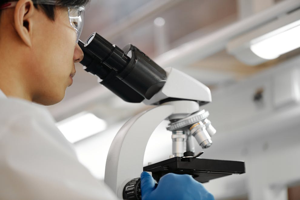
The funding, from Wellcome, will enable researchers to develop bioimaging tools to visualise the fundamental biology of microscopic fungal pathogens, and to provide training for researchers at the forefront of these diseases.
The team brings together experts from the University of Exeter, the University of Edinburgh, and the University of Cape Town. They are all part of the Mycology Bioimaging Initiative, an international collaboration of researchers working to understand pathogenic fungi.
During the six and half year project, the Mycology Bioimaging Initiative will focus on fungal species identified by the World Health Organization as Priority Pathogens.
Dr Elizabeth Ballou from the University of Exeter’s Medical Research Council Centre for Medical Mycology is the Mycology Bioimaging Initiative team lead and said: "Fungi cause disease through the act of growing. Growing as invasive filaments, they damage tissue, and growing as single cells, they increase in number and spread. The bioimaging approaches enabled by this project will allow us to study the early events that allow growth, which will be essential to developing new therapeutics and diagnostics.”
Fungal pathogens infect 6.5 million people each year, but very little is known about how they cause disease. New fungal pathogens have also repeatedly emerged over the last two decades, meaning scientists urgently need fundamental research to allow improved diagnostics and help identify new drug targets.
The goals of the Mycology Bioimaging Initiative are twofold: to develop specialist bioimaging tools for these poorly understood species and to disseminate these tools to the global research community through focused training and researcher exchanges.
The team will focus initially on four species, developing bioimaging tools, including microfluidics, fluorescent reporters, and computational pipelines to allow scientific insight.
The invasive Mucorales species cause devastating mucormycosis, including an outbreak in 2021 among 40,000 patients suffering from Covid. The team will also develop tools for the drug-resistant species Candida glabrata, which causes bloodstream infections.
Professor Peter Swain from the University of Edinburgh said: “Candida glabrata is an increasingly important cause of infections, particularly in hospitals. We're developing new imaging to watch individual fungal cells as they respond to antifungal drugs in real time. We want to understand why some cells can tolerate treatment while others cannot.”
The Edinburgh team, also including Dr Ivan Clark and Dr Edward Wallace and based in the Centre for Engineering Biology at the School of Biological Sciences, will be developing engineering-inspired approaches to making subcellular events visible using fluorescent protein reporters and to watching the growth of fungal cells in microfluidic “traps”.
Emergomyces fungal pathogens were first reported in 2013 in South Africa and now cause skin and systemic infections across the globe. Professor Claire Hoving, of the University of Cape Town and the CMM AFRICA Unit, Institute of Infectious Disease and Molecular Medicine said: “Emergomyces are now recognised to cause cutaneous and systemic infections worldwide. Leveraging advanced imaging technologies—supported by locally embedded yet globally connected systems—helps ensure that expertise, diagnostic capacity, and tools remain within the regions most impacted by disease. This approach reduces delays in diagnosis, guides more effective treatment strategies, and ultimately improves health outcomes for vulnerable populations.”
Cryptococcal infections of the brain are a leading cause of HIV/AIDS-related death and disability globally, yet we still understand remarkably little about how this fungus damages the brain. Professor Rachael Dangarembizi of the University of Cape Town’s Neuroscience Institute and CMM AFRICA Unit said: “Using advanced light-sheet imaging, our team will develop new tools to visualise Cryptococcus within intact brains, giving us an unprecedented view of how the infection spreads and disrupts the brain during this fatal disease."
A key challenge for scientists has been the lack of tools for emerging pathogens, and lack of access to tools that have been developed. This award will resource Africa Mycology Bioimaging labs and scientists with sample preparation, image capture, and image analysis training. The Initiative will grow a network of workshop trainees who will also benefit from research exchanges between sites of fungal disease burden and sites of advanced bioimaging technologies.
Darren Thomson, who leads the Initiative’s training in applied Mycology Bioimaging, said: “We’re excited to see the network of bench biologists bring more context of the invasive pathogen to the network of clinical based scientists, who in turn, can help guide more disease relevance to the bench scientist's experimental approach.”
The MBI will run annual workshops in Mycology Bioimaging across sites to build this important network year-on-year.
Dr Ballou adds: "The team we've built for the MBI is a key strength. We have experts in imaging, data analysis, microfluidics, infection biology, fungal cell biology, engineering biology, and, importantly, open science and training. We are prioritising sharing the tools we develop through annual training workshops and researcher exchanges, and we're very excited to see the wider medical mycology community take up these technologies in their own research."